All human blood contains bacteria
The following article was originally published on Jeff Rense’s site at http://rense.com/general77/dblooder.htm
Alan Cantwell is a retired dermatologist. He is the author of The Cancer Microbe: The Hidden Killer in Cancer, AIDS, and Other Immune Diseases, and Four Women Against Cancer: Bacteria, Cancer and the Origin of Life, both published by Aries Rising Press and available from Amazon.com. Dr Cantwell died in 2021.
Tom Detwiler’s website is www.bloodmicrobe.org.
Bacteria are everywhere. Our mouths, throat, nose, ears all harbor germs. A few bacteria in the urine are considered normal; and fecal material is largely composed of bacteria. But what about the blood?
Under ‘normal’ conditions physicians generally believe human blood is ‘sterile’. The idea of bacteria living in the blood normally is largely considered medical heresy.
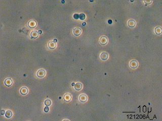
Recently Tom Detwiler of West Sayville, New York, sent me an email with three microphotographs he took from a video of a drop of his blood studied with ‘phase contrast’ and a ‘dark field microscope’. The photos clearly showed round and beaded forms emanating from red blood cells (erythrocytes), strongly suggesting the appearance of bacteria. (See Figures 1-3 below.) Detwiler is a biochemist with 18 years experience working as a microbiologist for a pharmaceutical company. He has an avid interest in dark field microscopy and microphotography. His blood findings were in accord with my own research documenting and photographing bacteria in many forms of cancer and other immune diseases.
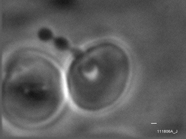
Figure 1. Erythrocyte (red blood cell) phenomenon. The release from the interior of the large red blood cell (in center) of tiny round bodies into the plasma. Phase contrast photo.
The idea that bacteria cause cancer is considered medical heresy. However, continuing research dating back to the late nineteenth century indicates that ‘pleomorphic’ (variably-appearing) bacteria are implicated in cancer. Over the past few decades more and more studies have confirmed that similar bacteria can be found in the blood.
Details of a century of research showing bacteria in cancer can be found in my two books The Cancer Microbe and Four Women Against Cancer. Although I personally have no experience with blood research and dark field microscopy, there are studies in the scientific literature that support Detwiler’s observations.
The evidence for blood bacteria
In a series of papers from 1972-1979, the late Guido Tedeschi and his colleagues at the University of Camerino in Italy presented remarkable findings indicating universal infection of the blood with staphylococcus-like and streptococcal-like microbes.
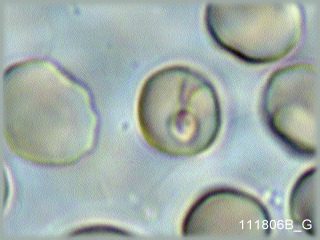
Figure 2. Erythrocyte phenomenon. The red blood cell in the center has a smaller round vacuole with moving bodies within. Phase contrast photo
In 1977 Domingue and Schlegel confirmed “the existence of a novel bacteriologic system” in the blood. They cultured staphylococcal-like bacteria and filamentous cocco-bacillary forms from 71% of the blood specimens from ill patients; and from 7% of supposedly healthy people. These pleomorphic bacteria grew out of round complex “dense bodies” and developed into “ordinary bacteria.” The authors concluded: “These organisms may represent an adaptation of certain bacteria to life in the blood.” Their full report, which contains pictures (full-screen) of the bacteria grown from blood, is online at http://www.pubmedcentral.nih.gov
In the 1990’s microbiologists Phyllis E Pease and Janice Tallak termed these blood bacteria as “the human bacterial endoparasite.” Finnish researchers Kajander et al describe them as “novel bacteria-like particles,” which are staphylococcal-like. Like viruses, these tiny bacterial forms were able to pass through bacterial filters, and were exceedingly difficult to culture. The Finnish team called them “nanobacteria” and proposed a tentative name for the novel agent: Nanobacterium sanquineum.
In 2002 McLaughlin et al presented a study entitled Are there naturally occurring pleomorphic bacteria in the blood of healthy humans? The researchers were surprised to discover bacteria in the blood “since it is generally acknowledged that the blood stream in healthy humans is a sterile environment, except when there is a breach in the integrity of the tissue membranes.”
Figure 3. Overview of the erythrocyte (red blood cell) phenomenon. Numerous small buds emanating from the red blood cells are visible, as well as smaller unattached darkly-colored buds in the surrounding solution.
A few critics claim that Detwiler’s forms are contaminating bacteria or ‘artifacts’ that are not microbial in origin. However, in view of recent studies, it is clear that bacteria do exist in human blood. Furthermore, bacteria are large enough to be observed microscopically. Thus, Detwiler’s observation of bacteria appears credible.
Bechamp, Enderlein, and Reich
In actuality, the study of the blood and the microbes that emanate from blood cells was the subject of extensive examination in the late nineteenth century by Antoine Bechamp (1816-1908). At the time, it was widely believed that the cell was the smallest unit of life. But the French professor insisted it was the tiny granules within the cell (which he called “microzymas”) which comprised the smallest unit of life. In Bechamp’s heretical view, bacteria could develop from these microzymas under appropriate conditions. His book, The Blood and its Third Element, is published by A Distant Mirror.
German zoologist Gunther Enderlein (1872-1968) devoted many years to the dark field microscopic study of the blood. The complicated “life cycle” of these blood bacteria is described in his book Bacterien-Cyclogenie (1925).
A controversial blood test is named after Enderlein; and in 1993 a bi-lingual German and English translation of his research was published entitled: Blood Examination in Darkfield: According to Prof. Dr. Gunther Enderlein. The book is heavily illustrated with color photos of bacteria in the blood. Although it is a difficult read due to Enderlein’s complex terminology of the various pleomorphic blood forms, it is considered an essential work for practitioners performing the highly controversial ‘live blood cell analysis’ of human blood. Enderlein believed that the sterility of the blood was an invalid assumption on the part of medical science. He claimed the blood elements of all vertebrates, up to and including man- even the healthiest-have been subjected to a massive infestation of primitive-phase ‘endobionts’.
The infection of the blood by bacteria is commonly accepted as fact by some alternative medical practitioners. However, attempts to make a medical diagnosis by dark field examination of the patient’s blood is considered a scam and “sheer hokum” by most medical doctors. A highly critical review of this procedure entitled Live blood cell analysis; Another gimmick to sell you something, by Stephen Barrett MD, can be found on quackwatch.org.
Other controversial researchers who made outstanding contributions to the study of pleomorphic microbes in human disease include Raymond Royal Rife, Wilhelm Reich and others. (For details of the scientific achievements of Royal Rife, see rife.org)
Also see:
- Synthesis of the work of Enderlein, Bechamp, and other pleomorphic researchers, by Dr Karl Poehlman
- Royal Raymond Rife by Jeff Rense at http://www.rense.com/health/rife.htm
- Dr. Wilhelm Reich: Scientific Genius or Medical Madman? by Alan Cantwell at http://whale.to/a/cantwell.html
In addition, the exhaustive and highly controversial ‘somatid’ work of Quebec biologist Gaston Naessens should be noted. Details of his research can be found on the Internet.
Pitfalls in the microbiology of the blood
The microbiology of the blood is intimately related to the proposed bacterial cause of cancer. The highly controversial microbiology of cancer was fully explored during the 1950s, 60s, and 70s by four largely ignored women scientists, namely Virginia Livingston MD, microbiologist Eleanor Alexander-Jackson PhD, cell cytologist Irene Diller PhD, and biochemist Florence Siebert PhD. These four remarkable scientists all recognized the extreme importance of bacteria in the blood. Details of their research appear in my book Four Women Against Cancer.
Much of the criticism against bacteria in cancer and in human blood revolves around the inability of scientists to precisely identify the species and/or multiple species of bacteria involved in the process. Human blood is undoubtedly an aquarium for multiple kinds of bacteria, all intimately interacting with each other and presumably passing genetic material back and forth between each other (via “plasmids” and “bacteriophages”).
In 2001, a molecular study by Nikkari et al found bacterial DNA in the blood. The inconclusive report was titled Does blood of healthy subjects contain bacterial ribosomal DNA? The researchers were unsure of the origin of these bacterial genetic sequences. Not surprisingly, none of the published blood research cited in this present report was mentioned by Nikkari.
Further complicating the question of blood bacteria is the century-old unresolved controversy of bacterial monomorphism versus pleomorphism. Most microbiologists and doctors believe bacteria multiply by simply dividing in half (binary fission). But pleomorphists believe that the reproduction of bacteria is highly complex and involves various growth forms within the body that are not recognized and accepted by traditional science. It is not possible to study the microbiology of blood (and cancer) without a knowledge of bacterial pleomorphism.
Yet another stumbling block is the terminology used to describe the various bacterial forms seen in the blood and the tissue. Bacteria in the blood have been described by various researchers as mycoplasma, L-forms, cell wall deficient bacteria, nanobacteria, and a host of other confusing and often synonymous terms.
Blood bacteria are thought to be connected with the origin of life. Livingston (1906-1990) believed these microbes were responsible not only for the initiation of life, but also acted as terminators leading to death, admittedly a difficult concept for most people to consider. Wilhelm Reich (1897-1957) referred to bacteria emanating from energy-depleted cells as ‘T-bacilli’, the ‘T’ derived from the German word Tod, meaning death. He found T-bacilli in both healthy and sick individuals. However, in the blood of sick people they were more numerous. Reich devised a blood test to measure the vitality of blood. (For details, Google ‘Reich blood test’)
Detwiler feels that the demonstration of bacteria in normal blood is frightening to many people, who would prefer not to know such things. In addition, the idea might be scary for people who receive blood transfusions.
Is human blood sterile?
Although it may be comforting to believe that human blood is sterile, common sense indicates it isn’t. According to bloodbook.com, five to ten percent of the cases of HIV infection are transmitted worldwide through the transfusion of infected blood or tainted blood products. Other diseases that can be transmitted by transfusion include viral, hepatitis B and C, syphilis, malaria and Chagas’ disease. Each year bad transfusions cause an estimated 8 to 16 million hepatitis B virus infections, 2.3 to 5 million hepatitis C virus infections and 80,000 to 160,000 HIV infections.
Currently in the U.S. all blood donors are tested for HIV-1 and HIV-2, HTLV-1, hepatitis B and C, and syphilis. Excluded from donating blood are people with a history of IV drug abuse and hepatitis, and those with male homosexual activity since 1977. Blood is not tested for West Nile virus, nor for herpes viruses such as human herpes-8 virus, the virus causing Kaposi’s sarcoma.
Many blood banks encourage patients to donate their own blood prior to the scheduled date of an elective surgery, in order to minimize the possibility of transfer of viruses.
Blood bacteria and human disease
Despite a century of modern medicine we know little about the cause of cancer and the many chronic diseases that accompany old age. Heart and blood vessel disease (arteriosclerosis) are the most common causes of death in the elderly. Could blood bacteria contribute to the cellular changes in the heart and blood vessels?
It is said that if he lives long enough every man will develop prostate cancer. Thus, there must be something intrinsic in every man that causes this. Could it be the build-up of bacteria in the blood, coupled with declining cell vigor, as claimed by Reich? For new research pointing to the possible connection between bacteria and prostate cancer, go to http://www.rense.com/general67/four.htm
Dr. Virginia Livingston thought blood bacteria served as a way for Mother Nature to force old people off the planet in order to make more room for younger and healthier people.
Pleomorphic bacteria have a “life cycle” and so do we. We ourselves are “pleomorphic” in that we begin life as microscopic beings and grow to produce new life by mixing our genetic material with others. When we die, we hope to continue as ‘spirit’ with eternal life. In his experiments Wilhelm Reich was astonished to discover that it was impossible to destroy the smallest living forms of life.
The inability of modern medicine to recognize the reality and importance of blood bacteria is the great tragedy of modern science.
Hopefully, this communication and the intriguing photos by Tom Detwiler will encourage others to explore the evidence for bacteria in the blood – and the idea that these bacteria are connected with the origin of life itself.
Addendum: Tom Detwiler’s ‘Activation Method of the Blood’
Attached is the so-called activation procedure that I’ve been using. I’ve been working with variants of this procedure for a number of years. The results are striking, more viscerally effective than electron micrographs. I have long felt that if it was published 60 years ago we may not be in the current situation on this matter.
Section 1 is self explanatory.
Section 2 is recommended for more careful consideration of the matter.
1) Activation of a microbiological factor associated with the erythrocyte
Incubation of a blood sample in buffer solution demonstrates an association of the majority of erythrocytes with a microbiological factor. A procedure to activate this factor is a preparation of 25 uL freshly drawn blood mixed with 0.4 mL NaHPO4 [0.18M] pH 7; followed with incubation in a 50C water bath for 60 minutes. The result is viewed with phase contrast.
A blood preparation from a normal healthy individual will display the following phenomena:
1. Evolution of spicules from erythrocytes.
2. Budding of erythrocytes.
3. The release from vesicles within the erythrocyte of forms resembling cocci, often in chains.
4. Large vesicles containing motile particles within erythrocytes.
5. Motile particles appearing in the plasma solution.
Remarks
Similar results can be obtained over a pH range including 4.5 – 7.5. The lower end of the temperature range to evoke this is about 42C. Isotonic sodium citrate at pH 7 is also effective.
Several assumptions are utilized to facilitate this presentation; that the expressed microbiological forms from the blood of a healthy individual are predominantly that of one microorganism with a capability of different formats of expression.
The activation phenomena can be viewed as a destabilization of the homeostatic maintenance of a microorganism within the erythrocyte. This microorganism appears to be capable of exiting the cell in several forms.
The evolution of erythrocyte spicules/filaments is regarded as a biophysical property of the erythrocyte membrane.
A noticeable increase in particles can observed in slide preparations of whole blood over the course of several hours. The blood would appear to have an innate capability of generation of particles.
The results obtained by this procedure support conclusions reached by some previous investigators. It may be reasonable to assume that this phenomena has been viewed previously, and possibly interpreted in similar fashion.
2) Biological control
An autoclaved isotonic solution of sodium phosphate or sodium citrate will produce the activation of the erythrocyte associated microorganism as described. Attention to the possible biological content of the solution used is necessary to increase the confidence level concerning a particular observation, or in performing further work with this microbe.
Biological particles capable of proliferation will frequently demonstrate an ability to withstand an autoclave cycle. Reliance on autoclave processing for biological control allows the possibility of significant biological factors entering an experimental preparation through solutions or surfaces. As there are indications that this erythrocyte microorganism is integrated into the plasma response to biological factors, there is more than the concern of introducing life forms into a preparation containing a specific microbe to be considered.
The following approach was used to produce a sodium phosphate solution containing minimal biological factors.
Water was provided by distillation, by a system configured to minimize water droplet “carry over” with the steam vapor. The distillation system used was modified by the placement of a water droplet trap in the vapor path. Frequent maintenance of the distillation flask reduced the numbers of a particle life form appearing in the distillate to a level difficult to detect by dark field inspection (40x objective; LT 1000/mL).
Disodium phosphate has a desirable property of withstanding considerable heat without polymerizing into a polyphosphate. Sodium phosphate solution [0.18 M] was prepared from disodium phosphate that was previously brought to 230 C for 2 hours. The solution was then pH adjusted to 7 with 1N HCl, and used immediately.
Glassware was warmed at 230 C for 2 hours.
References
- Domingue GJ, Schlegel JU. Novel bacterial structures in human blood: cultural isolation. Infect Immun. 1977 Feb;15(2):621-7.
- Kajander EO, Tahvanainen E, Kuronen I and Ciftcioglu N.
- Comparison of Staphylococci and Novel Bacteria-Like Particles from Blood. Zbl. Bakt. Suppl. 26, 1994.
- McLaughlin RW, Vali H, Lau PC, Palfree RG, De Ciccio A, Sirois M, Ahmad D, Villemur R, Desrosiers M, Chan EC. Are there naturally occurring pleomorphic bacteria in the blood of healthy humans? J Clin Microbiol. 2002 Dec;40(12):4771-5.
- Nikkari S, McLaughlin IJ, Bi W, Dodge DE, Relman DA. Does blood of healthy subjects contain bacterial ribosomal DNA? J Clin Microbiol. 2001 May;39(5):1956-9.
- Pease PE, Tallack JE. A permanent endoparasite of man. 1. The silent zoogleal/symplasm/L-form phase. Microbios. 1990;64(260-261):173-80.
- Tedeschi GG, Di Iorio EE. Penetration and interaction with haemoglobin of corynebacteria-like microorganisms into erythrocytes in vitro. Experientia. 1979 Mar 15;35(3):330-2.
- Tedeschi GG, Bondi A, Paparelli M, Sprovieri G. Electron microscopical evidence of the evolution of corynebacteria-like microorganisms within human erythrocytes. Experientia. 1978 Apr 15;34(4):458-60.
- Tedeschi GG, Amici D, Sprovieri G, Vecchi A. Staphylococcus epidermidis in the circulating blood of normal and thrombocytopenic human subjects: immunological data. Experientia. 1976 Dec 15;32(12):1600-2.
- Tedeschi GG, Amici D. Mycoplasma-like microorganisms probably related to L forms of bacteria in the blood of healthy persons. Cultural, morphological and histochemical data. Ann Sclavo. 1972 Jul-Aug;14(4):430-42.