by Robert Folk, professor emeritus at the Department of Geological Sciences, University of Texas.
Summary
Nannobacteria are very small living creatures in the 0.05 to 0.2 micrometer range. They are enormously abundant in minerals and rocks, and probably run most of the earth’s surface chemistry. Although I conjecture that they form most of the world’s biomass, they remain “biota incognita” to the biological world as their genetic relationships, metabolism, and other characteristics remain to be investigated.
Introduction
Nannobacteria are dwarf forms of bacteria, mostly 0.05 to 0.2 micrometers, about one-tenth the diameter and 1/1000 the volume of ordinary bacteria. The word was first published as “nanobacteria” by Richard Y. Morita in 1988, but I used the spelling “nanno-” to conform with geological usage, e.g., “nannoplankton.” In the microbiological world, the minute forms are commonly called “ultramicrobacteria,” regarded as stressed or resting forms of big bacteria, and thought to be both rare and dormant. However in soils, sediments, minerals and rocks they are enormously abundant and may perhaps represent a novel life form intermediate between normal bacteria and viruses (which are around 0.01 to 0.02 micrometers). It is contended (Folk 1992, 1993b and other references cited herein) that they are responsible for a great deal of mineral precipitation as well as conversion of igneous minerals to soils, and the corrosion of metals.
Discovery
The important role of nannobacteria in the mineralogical world was discovered through dumb luck, idle curiosity and random reading. There was no LIFETIME RESEARCH PLAN or THIS CAN GET ME LOTS OF NATIONAL FUNDING idea involved. I was simply looking for a good excuse to continue doing field work in Italy because I loved the food and lifestyle, and hit upon the idea of working on the travertines of Rome (travertine is a whitish type of limestone, usually porous, formed in springs, lakes and streams, and has been used as building stone in Rome for 2000 years). Together with Professor Henry S. Chafetz of the University of Houston, I began work on the Italian travertines in 1979. In the course of this research it was discovered by chance that “normal-sized” bacteria, mainly sulfur-oxidizers, had played a very substantial role in precipitating this stone from the warm springs at Tivoli. Before this discovery neither Chafetz nor myself knew or cared anything about bacteria, as we were specialists in microscopic examination of limestones. In 1988, I returned to Italy to study the hot-spring travertines of Viterbo, about 50 km northwest of Rome. A new electron microscope with magnifications up to 100,000X began to reveal hordes of tiny bumps and balls. At first I passed them off as artifacts of sample preparation or laboratory contamination, as had every other scientist who had studied minerals and rocks with the scanning electron microscope (SEM). (One must be very careful to use only a 30-second gold coat on the specimen, or one can indeed produce nannobody-looking artifacts; see Folk and Lynch 1997). After a year of doubts, a little reading in Microbiology unearthed the fact that very small cells called “ultramicrobacteria” did in fact exist. With further SEM work, slowly the realization dawned that there really were entombed in minerals enormously abundant cells of this minute size (Figure 1), and in some examples the minerals seemed to be entirely made up of nannobacteria as closely packed as beans in a bag. Sometimes within a single crystal of mineral, part of the crystal would be crowded with nannobacteria and parts would be deserted, belying the idea of artifacts or “that’s the way minerals naturally dissolve.” Their occurrence in chains and grape-like clusters further attested to their true living status. For several years the research went forward without any assistance or external funding, much to the consternation and puzzled frowns of geological colleagues. My first oral presentation of the idea (Folk 1992) elicited mostly a stony silence.

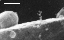
Figure 1(detail). Calcite from Bullicame hot springs, Viterbo, Italy; etched in dilute HCl. One elliptical “normal-sized” bacterial body is seen, about 1.5 micrometers long. Another normal-sized bacterium at right is only half emerged from its bed of calcite. The hordes of 0.1 micrometer spheres are nannobacteria; in the original SEM photo, the crystal appears to be almost wholly composed of nannobacteria. A small chain of about eight nannobacterial cells appears in the form of a homunculus in the center. A few spheroidal bodies, 0.2 to 0.6 micrometers, appear to bridge the size gap between ordinary bacteria and nannobacteria. Scale bar, 1 micrometer.
The idea of the existence of nannobacteria has been greeted with howls of disbelief by the majority of the biological community, who contend that these minute bodies cannot be bacteria because they are too small to contain the necessary genetic machinery for life. Even if they are not “normal” bacteria, they can easily be cultured (Figure 2) and those grown for a few days look exactly like those occurring in rocks and minerals (Figure 3) as old as 2 billion years or as recent as today, and are dead ringers for those occurring on Mars as to size, shape, and surface features (see McKay et al. 1996). Although DNA analysis of mineralized nannobacteria has yet to be done, some attempt has been made by medical researchers who find “nanobacterial” cells the same size as those I have observed, with cell walls that are very tough and that are resistant to acids, stains and poisons. Because of the tough walls special methods are required to isolate the DNA which occurs as very short strands (O. Kajander, Univ. Kuopio, Finland, personal communication).

Figure 2(detail). I placed a stub of metallic aluminum in Austin tap water for seven days. Gelatinous clots formed on the stub, and when viewed with the SEM these showed many diverse kinds of nannobacteria; the most noteworthy are the caterpillar-like bodies, about 0.05 micrometers wide, plus single bodies of the same size. These nannobacteria are perhaps metabolizing Al, and are good replicas of the Martian meteorite nannobacteria (McKay et al. 1996). Scale bar, 0.5 micrometers.

Figure 3(detail). Nannobacteria lying on top of a flake of chlorite clay; lava flow from Mt. Etna at Acireale, Sicily. These nannobacteria are involved in the precipitation of silicates (Lynch and Folk 1996). Compare these shapes with the nannobacteria grown on the Al stub in Figure 2, and with published Martian meteorite photos. Note single isolated nannobacteria about 0.03 micrometers, as well as the chains of partially merged bodies. Inorganic minerals do not crystallize like this. Scale bar, 0.5 micrometers.
Occurrence
At the initial discovery site, the hot springs of Viterbo, Lazio, Italy, some nannobacteria are found in untreated samples along with rare bacteria of normal size (Folk 1993b). However, upon slight etching with HCl, hordes of nannobacteria are revealed entombed in the calcite and aragonite crystals, like peanuts in peanut brittle (Figure 1). They occur in dense clumps and initiate precipitation; once the seed crystal has been formed, the remainder of the crystal continues to form abiotically (e.g., Pedone and Folk 1996). The nannobacteria act as catalysts, attracting cations to their negatively charged cell walls, leading to oversaturation on a submicrometer scale and triggering precipitation of CaCO3.
Once they were discovered in the travertines of Viterbo, nannobacteria were soon found in limestones and dolomites (CaMg(CO3)2) in rocks of all ages back to two billion years old (Folk 1993a). In some limestones they are abundant, in others they are scarce. Modern oolites of the Bahamas are rich in nannobacterially precipitated aragonite needles (Folk et al. 1996). Again, etching in dilute HCl is generally required to reveal them—more proof that they are not laboratory artifacts, but are contained within the rock. At Viterbo they are abundant in crystals of native sulfur (Folk 1992) and this fact led to their eventual discovery in metallic sulfide minerals such as pyrite, chalcopyrite and chalcocite (Sillitoe et al. 1996). Bacteria had long been suspected of playing a role in the deposition or beneficiation of ore minerals, but no one had looked for the presence of nannobacteria. In most sulfides they are revealed best by etching in HCl or HNO3.
Silica minerals also show evidence of precipitation by nannobacteria. Such has been observed to be the case with opal, chalcedony, chert and cristobalite (Folk et al. 1995). They are revealed by brief etching with HF. Again, some cherts consist of very closely packed nannobacteria, in other samples they are scanty or even absent.
Even complex silicates as clay minerals appear to have been produced by nannobacteria (Folk et al. 1994). The bacterially formed clays occur either as filaments with nannobacterial beads of the same size at their tips (like the head on a matchstick), as clumps of tiny balls or as swarms of caterpillar-like bodies (Figures 2 and 3). In clays the nannobacteria can range as small as 0.03 micrometers (Lynch and Folk 1996, Folk and Lynch 1997).
Preliminary studies imply that nannobacteria have an active role in the rusting of iron (Figure 4), the “greening” of copper, and the solubilization of metallic aluminum (Figure 2). They also contribute to the plugging of pipes by mineral “scaling,” and appear to be involved in the construction of the CaCO3 shells of clams, foraminifera, and even birds’ eggs.

Figure 4(detail). A piece of rusted iron from Port O’Conner, Texas. This was treated with HCl for 10 minutes to remove the iron hydroxide, revealing the naked nannobacterial cells that precipitated the iron. The cells cover the iron completely as a sheet of balls, there are a few short chains and one long filament composed of balls. The nannobacteria here mostly range from 0.02 to 0.06 micrometers. Scale bar, 0.5 micrometers.
Problems and Discussion
If nannobacteria are so abundant—possibly an order of magnitude more abundant than normal bacteria, if the abundance by volume in rocks can be indicative—how is it that they could have been missed for so long? Probably the main reason is that microbiologists have little or no interest in the occurrence of any type of bacteria in soils or rocks, and it has been standard microbiological dogma for fifty years that bacteria smaller than 0.2 micrometers cannot exist. This is the smallest size visible with an optical microscope, with which censuses for bacterial populations are taken, and filters of 0.2 micrometers are customarily used under the assumption that they will catch all the bacteria, and the fluid that runs through will be bacteria-free. The few geologists interested in the interaction between bacteria and minerals followed this dogma, and thus never looked at samples with the SEM at such high magnifications—and if they saw 0.1 micrometer bumps, they passed them off as laboratory contamination. You see what you are looking for and what you have faith in! A colleague once remarked that nannobacteria might be the “Dark Matter” of the Biological Universe—having enormous effects, but up to now not seen or recognized.
And so the study of nannobacteria chugged along at a sluggish pace until the electrifying discovery of the meteorite from Mars in Antarctica (McKay et al. 1996). Chris Romanek of NASA had heard my 1992 lecture, my “coming out of the closet” presentation on nannobacteria in carbonate rocks (Folk 1992) and decided to try the techniques on the Martian meteorite ALH84001. Forms in the 0.1 micrometer range, exactly resembling nannobacteria found on Earth, were discovered in this meteorite. The “incognoscenti” immediately attacked, using the arguments “too small to be bacteria,” “artifacts,” being unaware of the previous six years of work done on these minute creatures. In fact, unpublished photographs of work done by myself and F. Leo Lynch on Sicilian volcanic clays with nannobacteria can exactly duplicate every published photograph issued by NASA (Figure 3, Lynch and Folk 1996). Even more “far out,” work by us on a carbonaceous chondrite meteorite (Allende) reveals distinct grape-like clusters of 0.05 micrometer balls, probably nannobacteria as well, and this time kosher extraterrestrials without any chance of contamination from earthly ice or soils (Figure 5).

Figure 5(detail). A carbonaceous meteorite currently under study, collected on Feb. 8, 1969 from Allende, Mexico by meteoriticist Elbert King. Grape-like clusters of nannobacterial cells (size 0.02–0.10 micrometers), bridge a pore between two olivine crystals. I believe the majority of the carbon found within carbonaceous meteorites will be found to consist of these balls. Sample courtesy of D. Smith, Prof. of Geology, Univ. of Texas. Scale bar, 0.5 micrometers.
If the idea of the ubiquity and overwhelming biomass of nannobacteria on earth (and perhaps in space as well) is correct, this bears enormous implications for earth-surface chemistry, ore deposition, weathering, in microbiology and even medicine as a potential source of genes or hitherto unseen cause of diseases. Problems of environmental pollution, metal deterioration, and industrial processes should be heavily impacted. If the work on the Martian and other meteorites proves to be correct, extraterrestrial nannobacteria could perhaps be the “Dark Matter” of the astronomical universe as well, as Fred Hoyle and Chandra Wickramasinghe (1984) suggested some thirty years ago. Or maybe the whole idea is nuts, but believe me, the tiny particles do indeed exist, whatever form of quasibiont they represent, and some biologist needs to tell us what they really are.
References
Folk, R.L. 1992. Bacteria and nannobacteria revealed in hardgrounds, calcite cements, native sulfur, sulfide materials, and travertines (abstract). Geological Society of America Annual Meeting, Program Abstracts, p 104.
Folk, R.L. 1993a. Dolomite and dwarf bacteria (nannobacteria) (abstract). Geological Society of America Annual Meeting, Program Abstracts, A–397.
Folk, R.L. 1993b. SEM imaging of bacteria and nannobacteria in carbonate sediments and rocks. J. Sediment. Petrol. 63:990–999.
Folk, R.L. and F.L. Lynch. 1997. The possible role of nannobacteria (dwarf bacteria) in clay mineral diagenesis and the importance of careful sample preparation in high magnification SEM study. J. Sediment. Res. In press.
Folk, R.L., F.L. Lynch and E.T. Rasbury. 1994. Evidence for bacterial precipitation of clay minerals upon sand grains, in soils, and in the subsurface (abstract). Geological Society of America Annual Meeting, Program Abstracts, A–508.
Folk, R.L., P.J. Noble, G. Gelato and R.J.C. McLean. 1995. Precipitation of opal–CT lepispheres, chalcedony and chert nodules by nannobacteria (dwarf bacteria) (abstract). Geological Society of America Annual Meeting, Program Abstracts, A–305.
Folk, R.L., F.L. Lynch and R.P. Major. 1996. The role of nannobacteria (dwarf bacteria) in the formation of oolites and hardgrounds, Joulters Cays, Bahamas (abstract). Geological Society of America Annual Meeting, Program Abstracts, A–275.
Lynch, F.L. and R.L. Folk. 1996. Possible nannobacterial mediation in the alteration of volcanic rocks to clay minerals in South Italy (abstract). Geological Society of America Annual Meeting, Program Abstracts, A–90.
McKay, D.S., E.K. Gibson, K.L. Thomas-Keprta, L.H. Vali, C.S. Romanek, S.J. Clemett, Z.D.F. Chillier, C.R. Maechling and R.N. Zare. 1996. Search for past life on Mars: possible relic biogenic activity in Martian meteorite ALH84001. Science 273:924–926.
Morita, R.Y. 1988. Bioavailability of energy and starvation survival in nature. Can. J. Microbiol. 34:436–441.
Pedone, V.A. and R.L. Folk. 1996. Formation of aragonite cement by nannobacteria in the Great Salt Lake, Utah. Geology 24:763–765.
Sillitoe, R.H., R.L. Folk and N. Saric. 1996. Bacteria as mediators of copper sulfide enrichment during weathering. Science 272:1153–1155